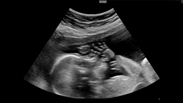
Ultrasound schedule during pregnancy
There are no rigid rules on the number of ultrasounds during pregnancy. Additional scanning must be done if there is a suspicion that a fetus has some abnormalities. Ultrasonography is recognized as the safest and most informative method of intrauterine examination. Read about benefits and risks of having frequent ultrasounds during pregnancy.

Pregnancy Ultrasound Schedule
There is a specific ultrasound schedule. Learn the table below.
| Timeframe | Objectives | compulsory | desirable | acc. to medical indications |
|---|---|---|---|---|
| 7th week | · confirmation of pregnancy · diagnostics of ectopic or molar pregnancy · confirmation of cardiac pulsation · measuring the length of the fetus from crown to rump · dating pregnancy · scanning the current status of the fetus · determining the location of ovum attachment | + | ||
| 11th — 12 thweesks | · diagnosis of Down syndrome by measuring the collar space of a fetus and evaluation of the nasal bone · scanning the structure of the brain · measuring the size of the brain ventricles · scanning the spine, arms and legs of the fetus, its internal organs | + | ||
| 1st trimester | Detection of diseases of the pelvic organs in an expectant mother (fibroids, cysts or tumors of the ovaries, uterus bicornis, intrauterine septum etc.) | + | ||
| 22nd week | · detection of congenital malformations · accurate survey of the anatomy of the baby, · detection of any heart malformations · identification of the lag in development · scanning the position of the placenta · prediction of a baby’s gender. | + | ||
| 32nd week | · baby size estimation · assessment of fetal growth, · screening for possible violations · amniotic fluid quantity estimation · detection of the placenta location · scanning of a baby position · estimation of the current state of the placenta · estimation of the stage of maturity of baby internal organs | + | ||
| 39th — 40th week | · estimation of baby size and in utero position before childbirth · detection of possible umbilical cord entanglement | + |
It’s a good idea to have at least 3 scheduled ultrasounds during pregnancy — at 12, 22 and 32 weeks.
There are certain symptoms, which clearly indicate possible pathology of pregnancy. The most common indications for performing additional ultrasound for pregnant women are:
- bleeding from the genital tract,
- pain in the lower abdomen,
- abnormalities of placental attachment,
- discrepancy between the size of a baby and his age;
- suspected fetal death.
The main indications for sonography before the 12th week.
- Detection of intrauterine pregnancy, especially in cases when there is a suspected ectopic pregnancy.
- Complications in early pregnancy, such as chorion detachment, non-developing pregnancy, complete or incomplete miscarriage.
- Examination to establish the presence or absence of cardiac activity of the fetus.
- Detection of clinically suspected multiple pregnancy.
You don’t need an ultrasound to confirm your pregnancy — use a pregnancy test, as you should try to avoid every possible influence on your fetus in the most vulnerable first trimester.


